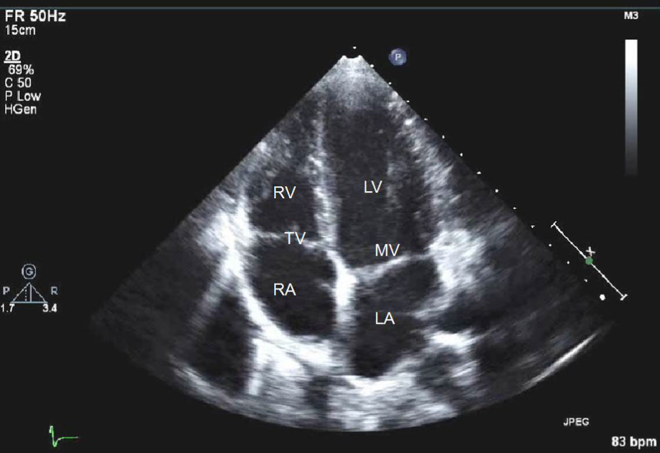
2D echo is the classic workhorse of heart imaging—simple, safe, and incredibly informative. It uses high-frequency sound waves to create real-time, two-dimensional images of your heart, allowing doctors to see the size, shape, and motion of the heart’s chambers and valves
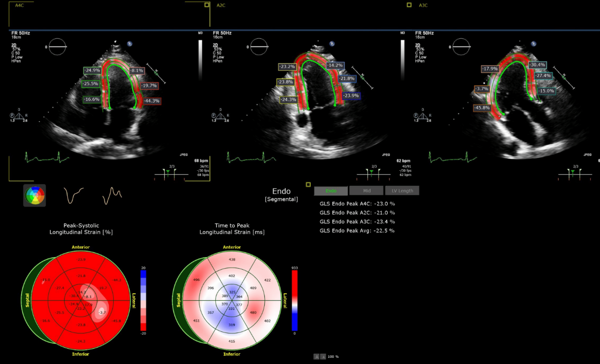
Here’s what it’s great for
Evaluating heart function: It shows how well your heart is pumping (ejection fraction).
Detecting valve problems: Like stenosis (narrowing) or regurgitation (leakage).
Spotting structural issues: Such as congenital heart defects or wall motion abnormalities.
Monitoring heart disease: Especially in conditions like cardiomyopathy or after a heart attack.
Strain Analysis
Strain analysis in echocardiography is a powerful technique used to assess how well the heart muscle deforms during contraction and relaxation.
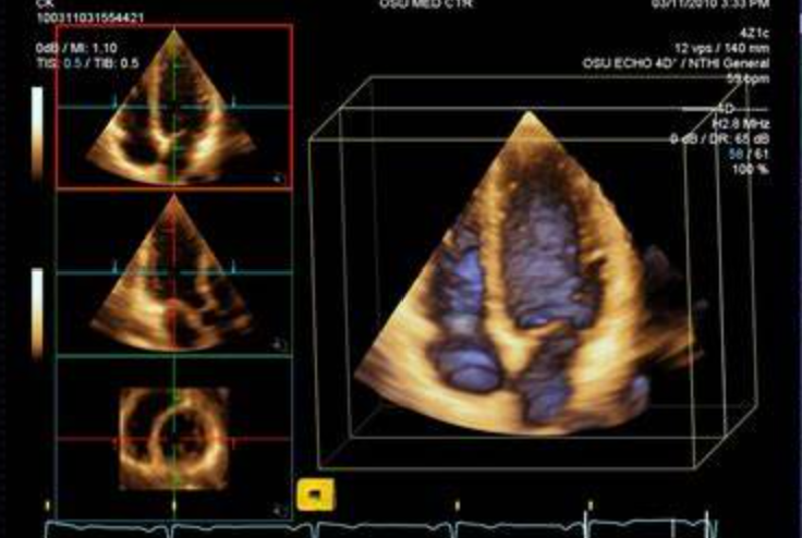
Strain is typically measured using speckle-tracking echocardiography (STE), which follows natural acoustic markers (speckles) in the heart muscle across frames
It evaluates myocardial deformation in three directions:
Longitudinal strain: shortening from base to apex (top to bottom).
Radial strain: thickening of the heart wall (inward motion).
Circumferential strain: squeezing around the heart’s circumference (like wringing a towel).
3D Echocardiography
3D echocardiography is like giving your heart a high-definition, three-dimensional photoshoot.
It builds on traditional 2D echo by capturing volumetric images of the heart, allowing clinicians to view structures from any angle in real time.
Here’s what makes it special:
Enhanced visualization: You can see heart valves, chambers, and congenital defects in stunning detail—especially useful for complex cases.
Accurate measurements: It provides more precise data on chamber volumes, ejection fraction, and valve morphology without relying on geometric assumptions.
Better assessment of valve diseases: Especially mitral and aortic valves—3D echo can show leaflet motion and regurgitant jets more clearly than 2D.